Edexcel A (SNAB) A Level Biology:复习笔记2.1.4 Cell Membranes

Cell Membranes
- Membranes are vital structures found in all cells
- The cell surface membrane creates an enclosed space separating the internal cell environment from the external environment, and intracellular membranes form compartments within the cell such as the nucleus, mitochondria, and endoplasmic reticulum
- Membranes do not only separate different areas but also control the exchange of substances from one side of a membrane to the other, as well as acting as an interface for communication
- Membranes are partially permeable
- Substances can cross membranes by diffusion and active transport
- Membranes contain receptor proteins, e.g. for binding to hormones, and antigens
- Membranes are partially permeable
Phospholipids
- Cellular membranes are formed from a double layer, or bilayer, of phospholipids
- Phospholipids consist of
- A molecule of glycerol
- A phosphate group, which forms the phosphate head
- Two fatty acid tails, making up the lipid tail
- Phospholipids contain two distinct regions: a polar head and two non-polar tails
- The phosphate head of a phospholipid is polar, meaning that it can interact with polar water molecules; the head is therefore described as being hydrophilic
- Hydro = water
- Philic = loving
- The lipid tail is non-polar, meaning that it cannot interact with polar molecules; the tail is therefore described as hydrophobic
- Hydro = water
- Phobic = hating
- The phosphate head of a phospholipid is polar, meaning that it can interact with polar water molecules; the head is therefore described as being hydrophilic
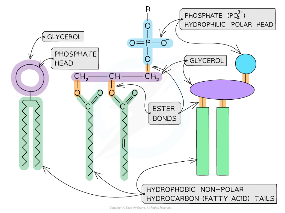
Phospholipids can be visually represented in different ways. In the image above the left-hand diagram shows a simple representation of the phosphate head and lipid tails, while the central diagram shows the chemical structure of each region; the separate glycerol and phosphate group can be seen, as well as the ester bonds that join the components together. The right-hand diagram shows a diagrammatic representation of the chemical structure.
- If phospholipids are spread over the surface of water they form a single layer with the hydrophilic phosphate heads in the water and the hydrophobic fatty acid tails sticking up away from the water
- This is called a phospholipid monolayer
- Alternatively, two-layered structures may form in sheets; these are called phospholipid bilayers
- Phospholipid bilayers form the basic structure of the cell membrane
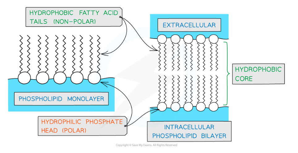
Phospholipids can form monolayers and bilayers
Structure of membranes
- The phospholipid bilayers that make up cell membranes also contain non-lipid components
- Proteins are involved with cell transport and communication
- The proteins can either be intrinsic or extrinsic
- Intrinsic proteins can also be referred to as integral
- Extrinsic proteins can also be referred to as peripheral
- Intrinsic proteins are embedded in the membrane with their precise arrangement determined by their hydrophilic and hydrophobic regions
- Extrinsic proteins are found on the outer or inner surface of the membrane
- The proteins can either be intrinsic or extrinsic
- Cholesterol can be found between the phospholipids, where it regulates membrane fluidity
- Cholesterol increases the fluidity of the membrane at low temperatures, stopping it from becoming too rigid
- This occurs because cholesterol stops the phospholipid tails packing too closely together
- Interaction between cholesterol and phospholipid tails also stabilises the cell membrane at higher temperatures by stopping the membrane from becoming too fluid
- Cholesterol molecules bind to the hydrophobic tails of phospholipids, stabilising them and causing phospholipids to pack more closely together
- Cholesterol increases the mechanical strength and stability of membranes; without it membranes would break down and cells would burst
- Cholesterol increases the fluidity of the membrane at low temperatures, stopping it from becoming too rigid
- Glycolipids and glycoproteins are present on the surface of the cell, where they aid cell-to-cell communication
- Glycoproteins are proteins with carbohydrate attached, while glycolipids are lipids with carbohydrate attached
- The glycolipids and glycoproteins bind with substances at the cell’s surface, e.g. hormones
- Some glycolipids and glycoproteins act as cell markers or antigens for cell-to-cell recognition
- E.g. the ABO blood group antigens are glycolipids and glycoproteins that differ slightly in their carbohydrate chains
- Proteins are involved with cell transport and communication
- The phospholipid bilayer with its additional components is often described as a 'fluid mosaic'
- The scattered pattern produced by the components within the phospholipid bilayer looks somewhat like a mosaic when viewed from above
- The mosaic of phospholipids and proteins can move around within the bilayer by diffusion, hence the mosaic is said to be 'fluid'
- The phospholipids mainly move sideways, within their own layer
- The many different types of proteins interspersed throughout the bilayer move about within it, although some may be fixed in position
- Note that the fluid mosaic model is one model of membrane structure; other models have been considered and rejected as knowledge of membrane structure has advanced
- The membrane is partially permeable
- Small, non-polar molecules can pass through the gaps between the phospholipids
- Large, polar molecules must pass through specialised membrane proteins called channel proteins and carrier proteins
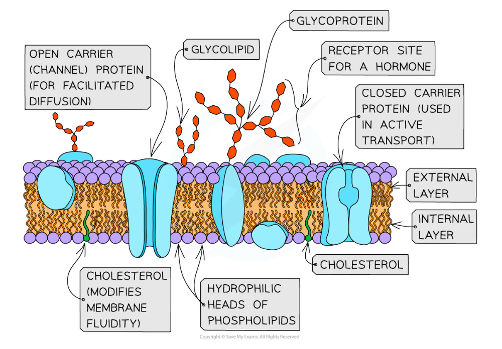
The distribution of the proteins within the membrane gives a mosaic appearance and the structure of proteins determines their position in the membrane
Exam Tip
In an exam it is good practice to always refer to the cell membrane that surrounds the cell as either the cell surface membrane or the plasma membrane; this distinguishes it from all of a cell's internal membranes
Models of the Cell Membrane
- Scientists use models to represent real world ideas, organisms, processes, and systems that cannot be easily investigated
- Over time, as technological developments have been made, the models used to represent the structure of cell membranes have changed
- The fluid mosaic model of cell membrane structure was first proposed in 1972 by Singer and Nicolson
- The model has evolved over time, and is thought to best account for the structure and functions of cell membranes as scientists currently understand them
- Models can change on the basis of new discoveries; if scientists found a new feature of cell membranes that didn't fit with the fluid mosaic model, the model would be altered, or a new model introduced
- Evolving cell membrane models have included the following
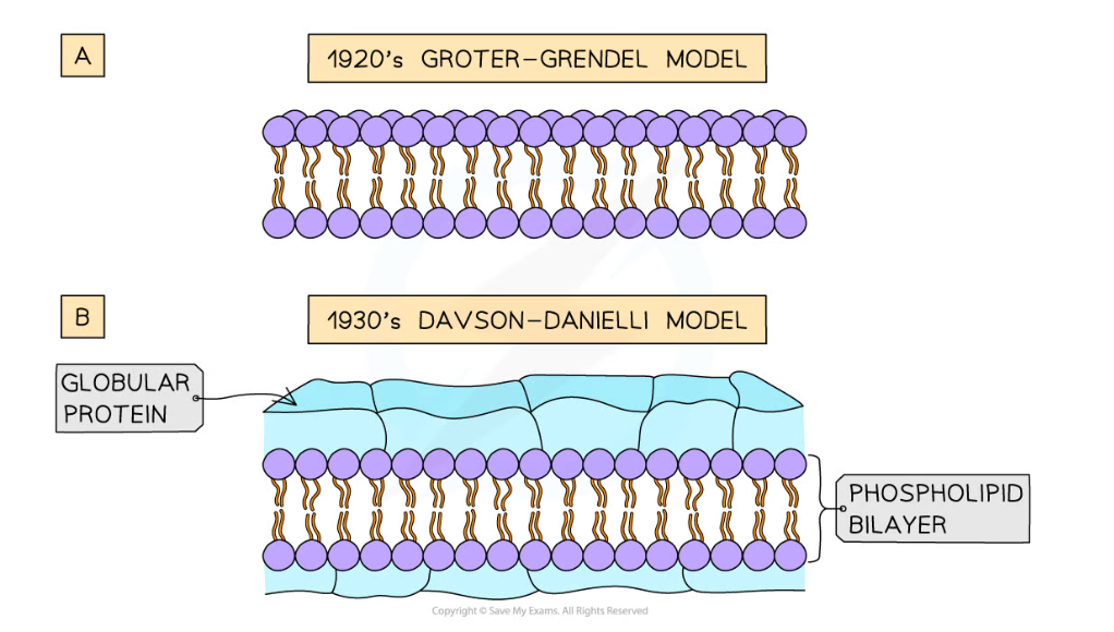
- The Gorter and Grendel model of the 1920s showed that the phospholipids in the membrane of cells were arranged into a bilayer
- Evidence for this model
- The number of phospholipids extracted from red blood cell membranes was double the area of the plasma membrane if it was arranged as a monolayer
- Problems with this model
- Their model did not explain the location of proteins or how molecules that were insoluble in lipids moved into and out of the cell
- Evidence for this model
- Davson and Danielli's model of the membrane from the 1930s suggested that the proteins were arranged in layers above and below the phospholipid bilayer
- Evidence for this model
- Membranes were effective at controlling the movement of substances in and out of cells
- Electron micrographs showed the membrane had two dark lines with a lighter band between
- In electron micrographs, proteins appear darker than phospholipids
- Problems with this model
- Freeze etched electron micrographs of the centre of the membrane showed globular structures scattered throughout
- Improvements in technology used to analyse the proteins in the membranes showed that proteins were globular, varied in size and had parts that were hydrophobic
- Evidence for this model
- Singer and Nicolson proposed the fluid mosaic model in the 1970s; the model stated that membranes were fluid and that the globular proteins were both peripheral and integral
- Evidence for this model
- Analysis of freeze-etched electron micrographs showed proteins extending into the centre of membranes
- Biochemical analysis of the plasma membrane components showed that membrane proteins are free to move within the bilayer
- Evidence for this model
- The Gorter and Grendel model of the 1920s showed that the phospholipids in the membrane of cells were arranged into a bilayer
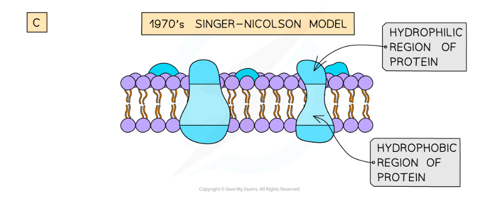
Models of cell membrane structure have evolved over time
Exam Tip
You do not need to recall the names of the scientists or dates for different models of membrane structure, but it is important to understand that models of membrane structure are interpretations of data which can change when scientific advances enable new discoveries.
Remember that models represent real-life structures and processes.
转载自savemyexams
以上就是关于【Edexcel A (SNAB) A Level Biology:复习笔记2.1.4 Cell Membranes】的解答,如需了解学校/赛事/课程动态,可至翰林教育官网获取更多信息。
往期文章阅读推荐:
全网破防!ALevel CIE数学M1疑似错题?经济P2难度飙升?5月6日大考考情分析必看!
A-Level CIE就大规模泄题发布最严处罚!哪些考生必须重考?你的成绩怎么办?

翰林AMC8视频课重磅上线!

国际竞赛真题资源免费领取