CIE A Level Biology复习笔记7.1.4 Phloem Sieve Tube Elements

Phloem Sieve Tube Elements & Companion Cells: Structure & Function
- The function of phloem tissue in a plant is to:
- Transport organic compounds (assimilates), particularly sucrose, from the source (eg. leaf) to the sink (eg. roots). The transport of these compounds can occur up and down the plant
- The organic compounds are dissolved in water to form sap
- Phloem is a complex tissue made up of various cell types; its bulk is made up of sieve tube elements which are the main conducting cells and companion cells
- Other cell types of phloem tissue also include parenchyma for storage and strengthening fibres
- Mature phloem tissue contains living cells, unlike xylem tissue
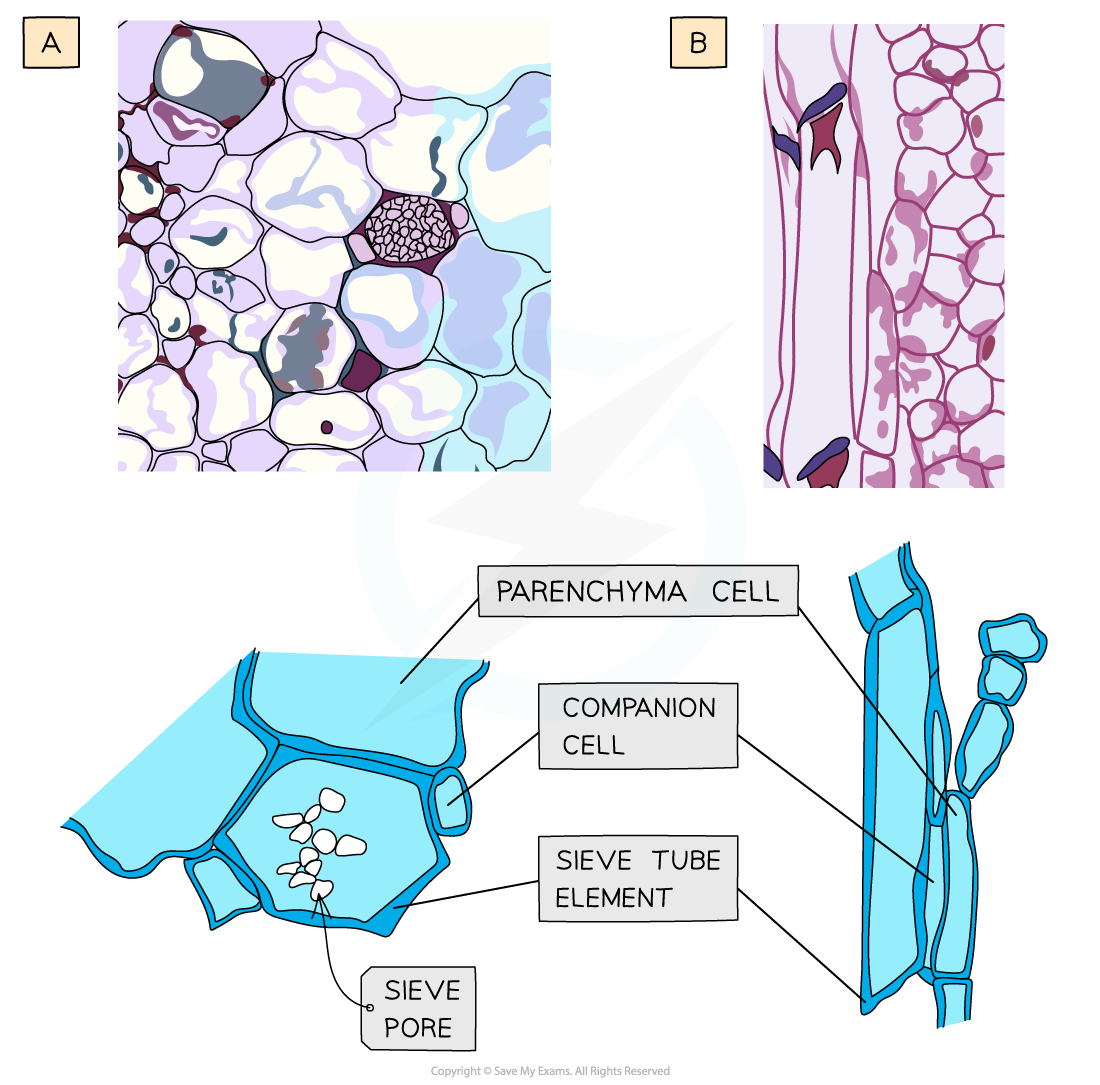
Structure of phloem tissue. (a) Microscope slide image and drawing of a sieve tube element and companion cell in transverse section (TS), (b) photomicrograph image and drawing of a sieve tube element and companion cell in longitudinal section (LS).
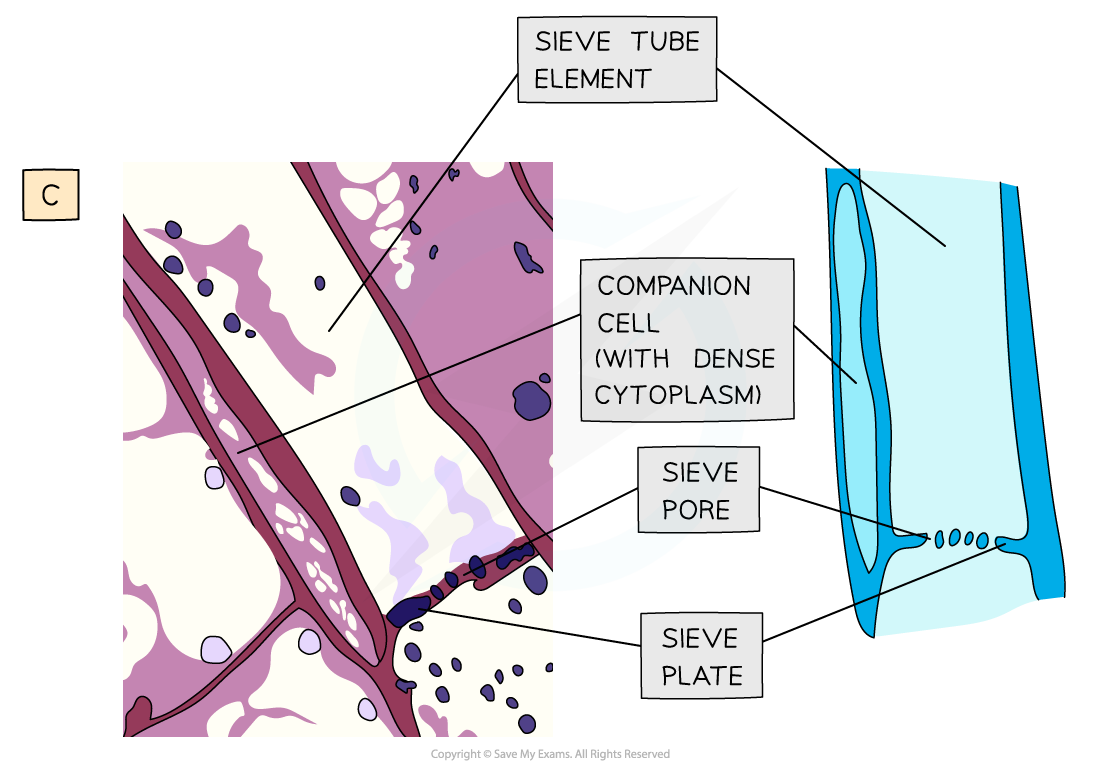
Structure of phloem tissue. (c) Transmission electron micrograph image and drawing of a sieve tube element and companion cell in transverse section (TS)
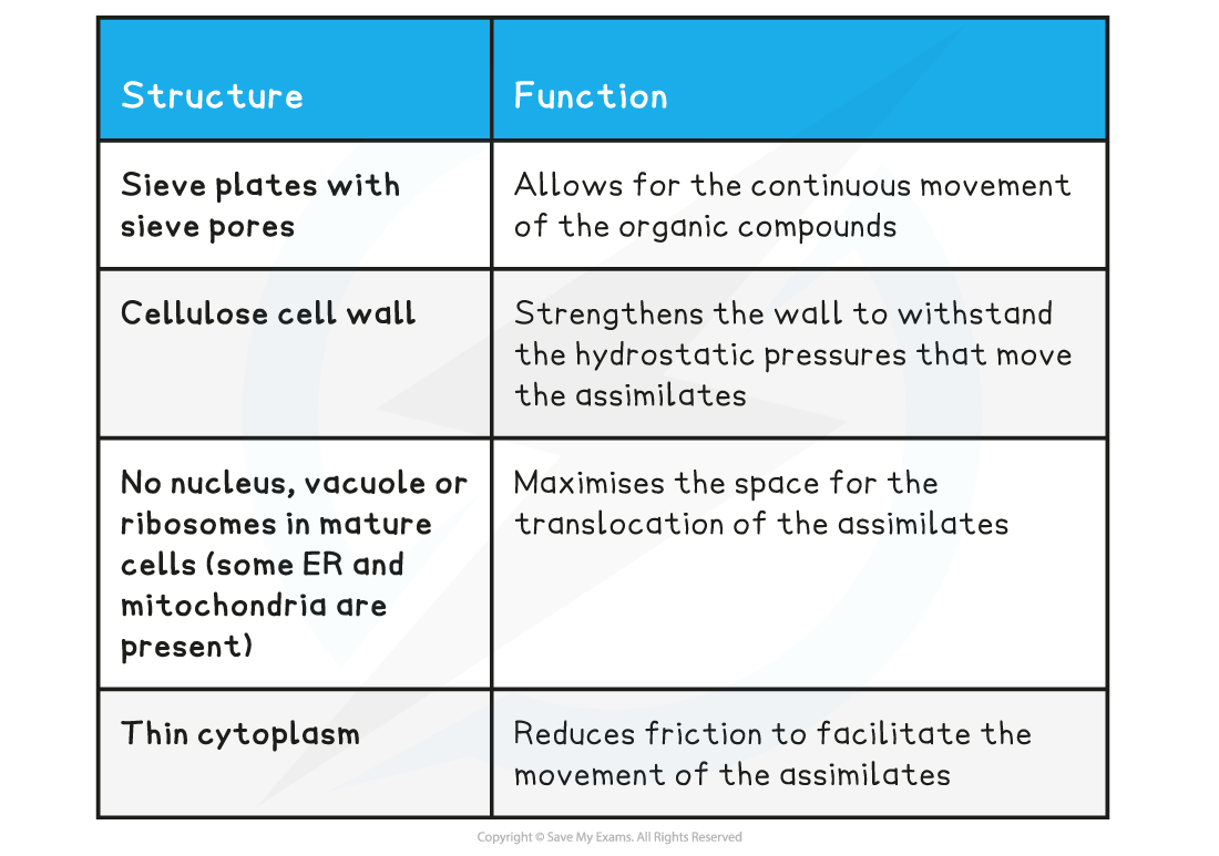
Sieve tube elements
- Sieve tube elements line up end to end to form a continuous tube
Phloem sieve tube elements structure & function table
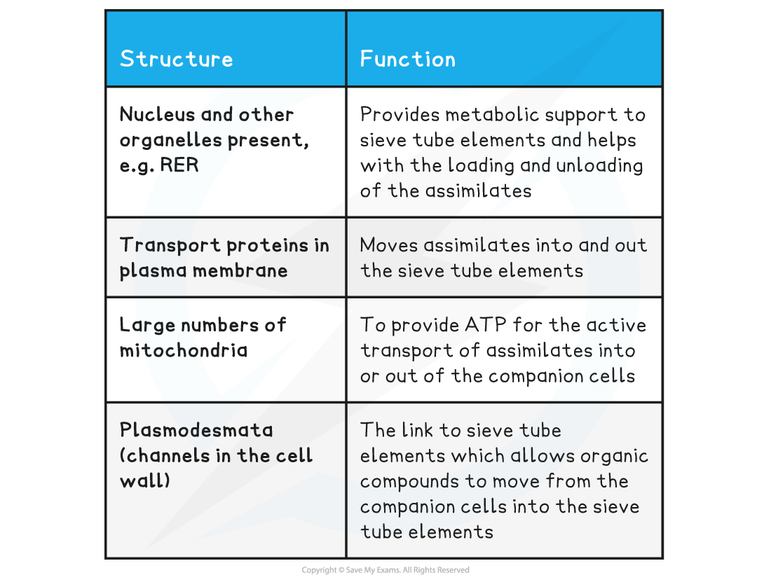
Companion cells
- Each sieve tube element has a companion cell associated with it as companion cells control the metabolism of their associated sieve tube member
- They also play a role in loading and unloading of sugars into the phloem
Phloem companion cells structure & function table
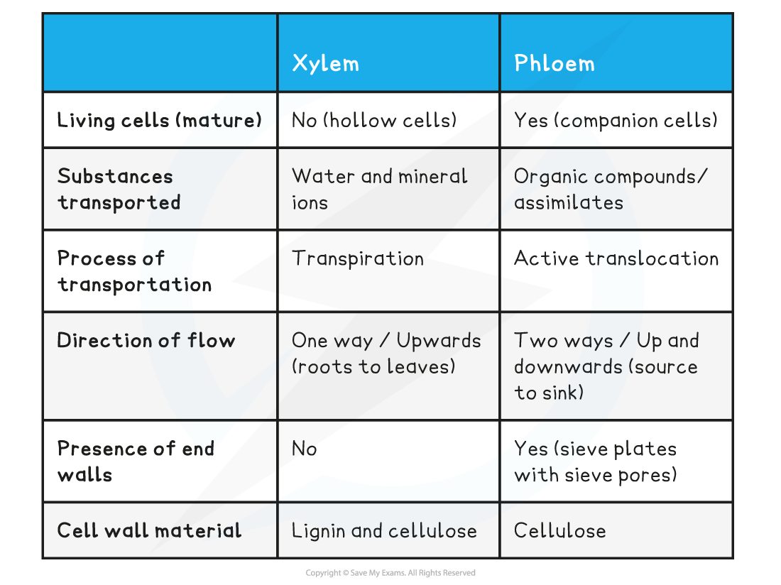
Comparison of xylem & phloem tissue table
Exam Tip
Understand the difference between sieve tube elements and companion cells, and how they are different to xylem tissue. Remember that mature xylem tissue is dead, so there is no evidence of organelles, and they have lignified cell walls, whereas sieve tube elements have no lignin, do have sieve plates, and their companion cells contain nuclei and dense cytoplasm.
转载自savemyexams
以上就是关于【CIE A Level Biology复习笔记7.1.4 Phloem Sieve Tube Elements】的解答,如需了解学校/赛事/课程动态,可至翰林教育官网获取更多信息。
往期文章阅读推荐:
全网破防!ALevel CIE数学M1疑似错题?经济P2难度飙升?5月6日大考考情分析必看!
A-Level CIE就大规模泄题发布最严处罚!哪些考生必须重考?你的成绩怎么办?

翰林AMC8视频课重磅上线!

国际竞赛真题资源免费领取