IB DP Biology: SL复习笔记1.3.6 Skills: Membrane Structure & Transport

Drawing the Fluid Mosaic Model
Membranes
- Membranes are vital structures found in all cells
- The cell surface membrane creates an enclosed space separating the internal cell environment from the external environment
- Intracellular membranes (internal membranes) form compartments within the cell, such as organelles (including the nucleus, mitochondria and RER) and vacuoles
- Membranes not only separate different areas but also control the exchange of materials passing through them; they are partially permeable
- Membranes form partially permeable barriers between the cell and its environment, between cytoplasm and organelles and also within organelles
- Substances can cross membranes by diffusion, facilitated diffusion, osmosis and active transport
- Membranes play a role in cell signaling by acting as an interface for communication between cells
Membranes formed from phospholipid bilayers help to compartmentalise different regions within the cell, as well as forming the cell surface membrane
Fluid Mosaic Model
- The fluid mosaic model of membranes was first outlined in 1972 by Singer and Nicolson and it explains how biological molecules are arranged to form cell membranes
- The fluid mosaic model also helps to explain:
- Passive and active movement between cells and their surroundings
- Cell-to-cell interactions
- Cell signalling
- The fluid mosaic model describes cell membranes as ‘fluid’ because:
- The phospholipids and proteins can move around via diffusion
- The phospholipids mainly move sideways, within their own layers
- The many different types of proteins interspersed throughout the bilayer move about within it (a bit like icebergs in the sea) although some may be fixed in position
- The fluid mosaic model describes cell membranes as ‘mosaics’ because:
- The scattered pattern produced by the proteins within the phospholipid bilayer looks somewhat like a mosaic when viewed from above
- The fluid mosaic model of membranes includes four main components:
- Phospholipids
- Cholesterol
- Glycoproteins and glycolipids
- Transport proteins
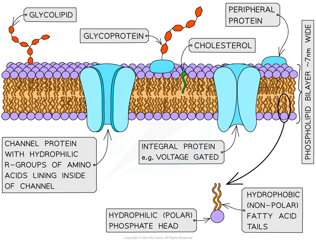
The main components of cell membranes. The distribution of the proteins within the membrane gives a mosaic appearance and the structure of the proteins determines their position in the membrane.
Exam Tip
When drawing the fluid mosaic model remember to include (and label) the phospholipid bilayer (making it clear which part is the phosphate head and which parts are the hydrocarbon tails), the thickness of the membrane (7 - 10 nm), integral proteins (show then embedded in the phospholipid bilayer and include a couple of different types e.g. channel/carrier), peripheral proteins (do not extend the protein into the hydrophobic region), glycoprotein (with a carbohydrate attached) and finally cholesterol (ensure the orientation is correct, OH group next to the phosphate heads and the rest positioned next to the tails).
Analysis of Evidence: Davson-Danielli Model
- Analysis of evidence from electron microscopy led to the proposal of the Davson-Danielli model
- Other methods were then used to further investigate the model and suggested evidence against the model
- Freeze-etchings
- Fluorescent markers of membrane proteins
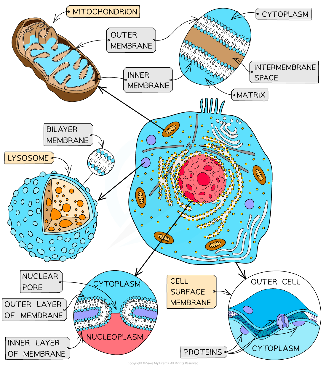
Transmission electron micrograph (TEM) of the plasma membrane
- When analysing transmission electron micrographs comment on:
- How the membrane has two darker layers surrounding a lighter line
- Proteins were known to appear darker in electron micrographs
- These were the observations that supported Davson-Danielli's model
TEM of a plasma membrane suggests evidence for Davson-Danielli's model
Freeze-etched electron micrographs
- When asked to analyse freeze-etched electron micrographs note that the very small bumps seen on the membranes are the integral proteins
- This provided evidence against Davson-Danielli’s model as it showed proteins extending into the centre of the membrane
- Be careful if the image is of a nuclear membrane as the larger circles represent the nuclear pores
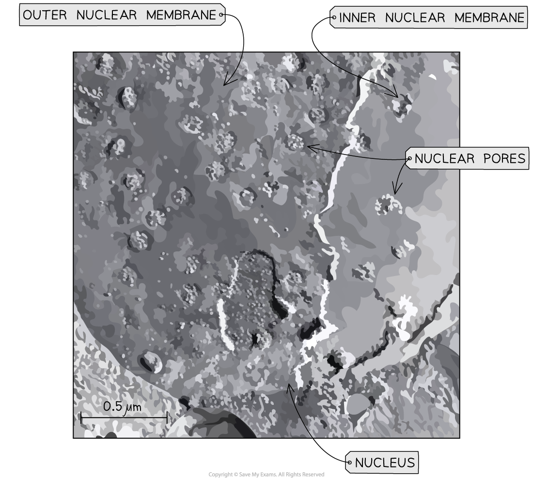
Freeze-etching of a nucleus suggests evidence against Davson-Danielli's model
Fluorescent markers on membrane proteins
- When analysing data on the use of red and green fluorescent markers attached to membrane proteins, the key evidence to note, is that as time progresses after the fusion of the different cells with the different markers has occurred, more mixing of the markers is observed
- This evidence did not support Davson-Danielli’s model that the proteins were a uniform layer above and below the phospholipids
- It supported the ‘fluid’ part of Singer & Nicolson’s fluid mosaic model as it suggested that membrane proteins can move
Fluorescent markers on membrane proteins suggest evidence against Davson-Danielli's model
Falsification of Davson-Danielli Model
NOS: Falsification of theories with one theory being superseded by another; evidence falsified the Davson-Danielli model
- For about 30 years the technology available to scientists supported the Davson-Danielli model of membrane structure
- From the 1950's an advancement in technology led to the accumulation of evidence which resulted in the Davson-Danielli model being superseded by the Singer-Nicolson 'fluid mosaic model'
- Analysis of freeze-etched electron micrographs showed proteins extending into the centre of membranes
- Biochemical analysis of membranes suggested that it was unlikely proteins formed continuous layers because it showed:
- Proteins were globular
- Varied in sizes
- They had parts that were hydrophobic
- The use of coloured fluorescent markers of antibodies showed that within forty minutes of fusing cells with different coloured fluorescent markers the markers had mixed
- This suggested that membrane proteins were free to move within the layer
Exam Tip
It is important to be able to provide the reasons why the evidence collected falsified the Davson-Danielli model.
转载自savemyexams
以上就是关于【IB DP Biology: SL复习笔记1.3.6 Skills: Membrane Structure & Transport】的解答,如需了解学校/赛事/课程动态,可至翰林教育官网获取更多信息。
往期文章阅读推荐:
IBO官宣2027年课改:数学、语言、艺术全调整,你的学习计划要更新了!
深耕九载!30+国际竞赛/课程讲义,硕博100%团队操刀,助力爬藤冲G5!

翰林AMC8视频课重磅上线!

国际竞赛真题资源免费领取