IB DP Biology: SL复习笔记1.2.4 Eukaryotic Cell Structure

Eukaryotic Cell Structure
Compartmentalized cell structure
- Eukaryotic cells have a more complex ultrastructure than prokaryotic cells
- The cytoplasm of eukaryotic cells is divided up into membrane-bound compartments called organelles. These compartments are either bound by a single or double membrane
- The compartmentalization of the cell is advantageous as it allows:
- Enzymes and substrates to be localised and therefore available at higher concentrations
- Damaging substances to be kept separated, e.g. digestive enzymes are stored in lysosomes so they do not digest the cell
- Optimal conditions to be maintained for certain processes e.g. optimal pH for digestive enzymes
- The numbers and location of organelles to be altered depending on requirements of the cell
- Eukaryotic cells have a key compartment called the nucleus
Animal and plant cells
- Animal and plant cells are both types of eukaryotic cells that share key structures such as:
- Membrane-bound organelles, including a nucleus
- Larger ribosomes (80S)
- However, there are key differences:
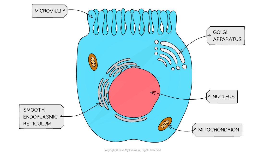
- Animal cells contain centrioles and microvilli
- Plant cells have a cellulose cell wall, large permanent vacuoles and chloroplast
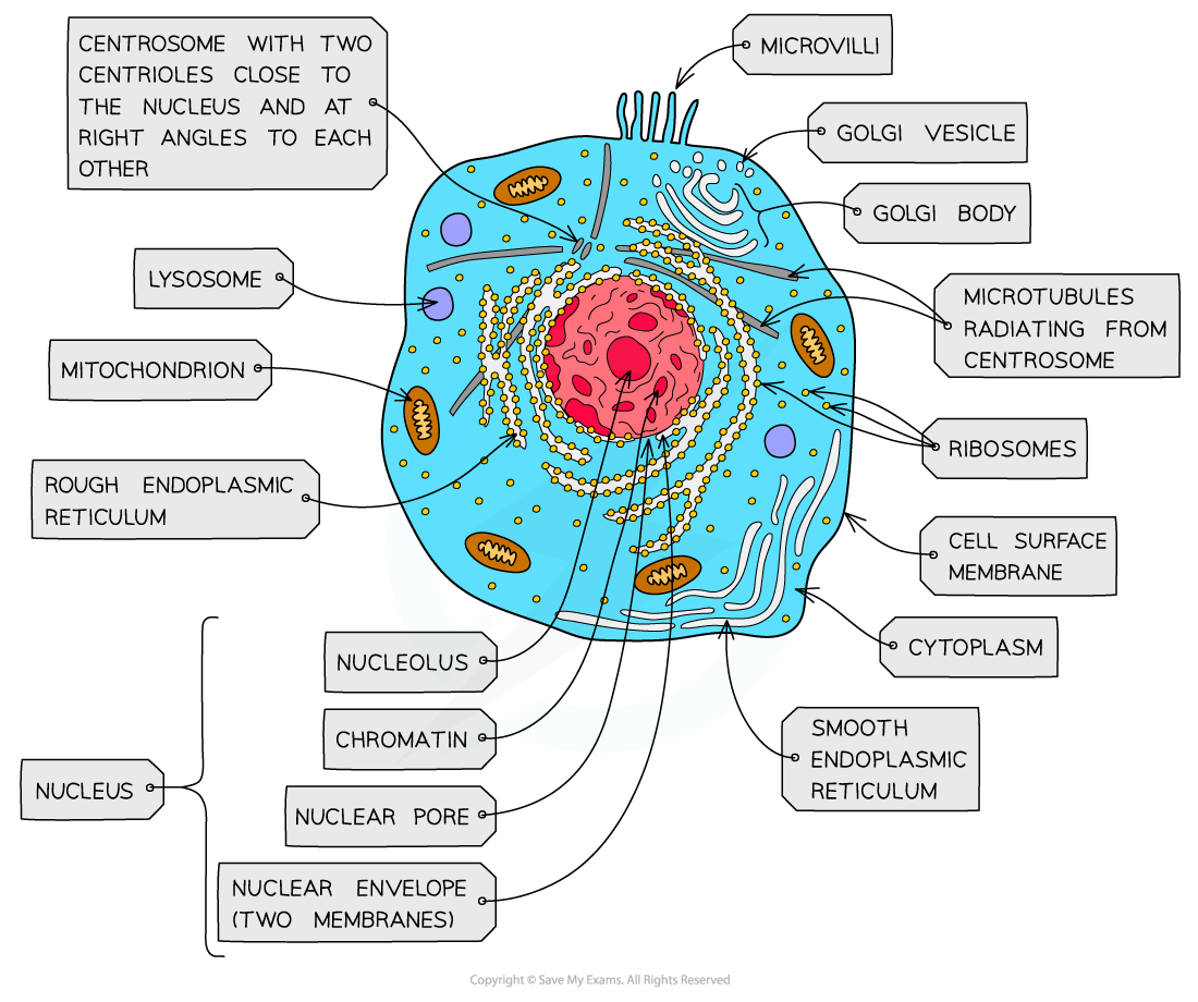
The ultrastructure of an animal cell shows a densely packed cell – the ER and RER and ribosomes form extensive networks throughout the cell in reality
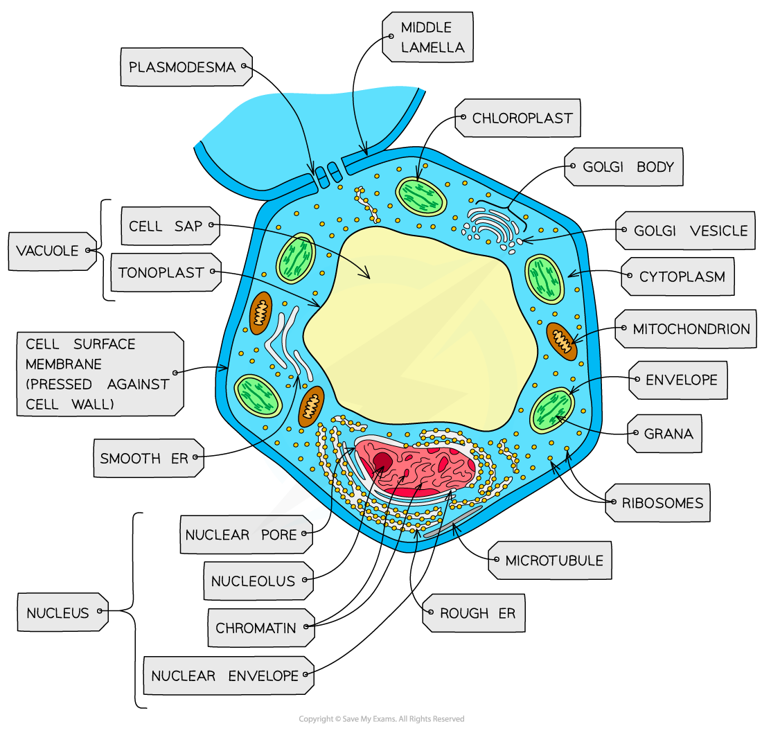
Plant cells have a larger, more regular structure in comparison to animal cells
- In complex multicellular organisms, eukaryotic cells become specialised for specific functions
- These specialised eukaryotic cells have specific adaptations to help them carry out their functions
- For example, the structure of a cell is adapted to help it carry out its function (this is why specialised eukaryotic cells can look extremely different from each other)
- Structural adaptations include:
- The shape of the cell
- The organelles the cell contains (or doesn’t contain)
- For example:
- Red blood cells are biconcave and do not contain a nucleus. This makes more space inside the cell so that they can transport as much oxygen as possible
- Cells that make large amounts of proteins will be adapted for this function by containing many ribosomes (the organelle responsible for protein production)
Organelles
Plasma membrane
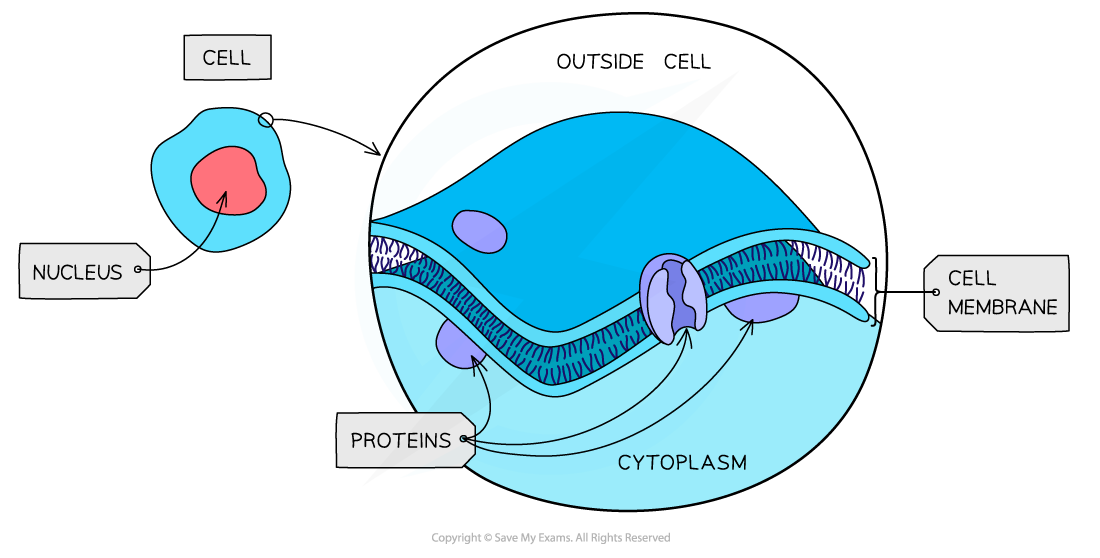
The structure of the cell surface membrane – although the structure looks static the phospholipids and proteins forming the bilayer are constantly in motion
- All cells are surrounded by a plasma membrane which controls the exchange of materials between the internal cell environment and the external environment
- The membrane is described as being ‘partially permeable’
- The plasma membrane is formed from a phospholipid bilayer of phospholipids spanning a diameter of around 10 nm
Nucleus
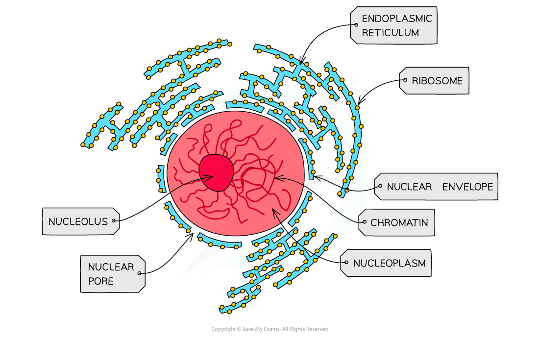
The nucleus of a cell contains chromatin (a complex of DNA and histone proteins) which is the genetic material of the cell
- Present in all eukaryotic cells (except red blood cells), the nucleus is relatively large and separated from the cytoplasm by a double membrane (the nuclear envelope) which has many pores
- Nuclear pores are important channels for allowing mRNA and ribosomes to travel out of the nucleus, as well as allowing enzymes (eg. DNA polymerases) and signalling molecules to travel in
- The nucleus contains chromatin (the material from which chromosomes are made)
- Chromosomes are made of sections of linear DNA tightly wound around proteins called histones
- Usually, at least one or more darkly stained regions can be observed – these regions are individually termed ‘nucleolus’ (plural: nucleoli) and are the sites of ribosome production
Rough endoplasmic reticulum
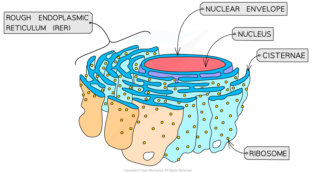
The rough endoplasmic reticulum (RER) - the attached ribosomes enable this structure to be identified in electron micrographs
- Found in plant and animal cells
- Surface covered in ribosomes (80S)
- Formed from continuous folds of membrane continuous with the nuclear envelope. These flattened membrane sacs are called cisternae
- Processes proteins made by the ribosomes
- The proteins synthesised by the ribosomes, move to the cisternae, bud off into vesicles that carry the proteins to Golgi apparatus before being secreted out of the cell
Ribosomes
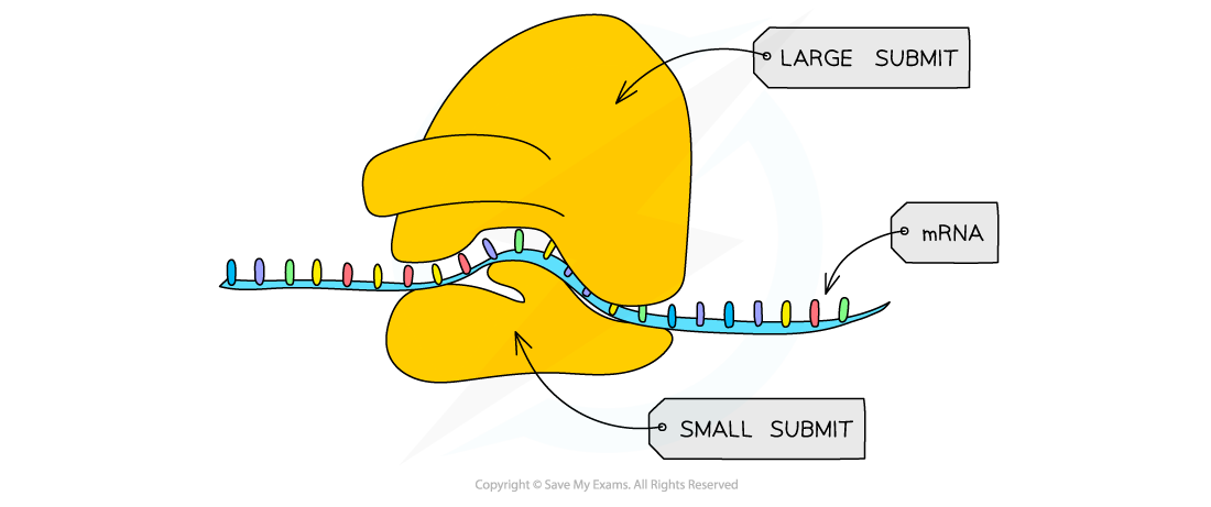
Ribosomes are formed in the nucleolus and are composed of almost equal amounts of RNA and protein
- Found freely in the cytoplasm of all cells or as part of the rough endoplasmic reticulum in eukaryotic cells
- Each ribosome is a complex of ribosomal RNA (rRNA) and proteins. They are constructed in the nucleolus (a region in the nucleus)
- 80S ribosomes (composed of 60S and 40S subunits) are found in eukaryotic cells
- Site of translation (protein synthesis)
Mitochondrion
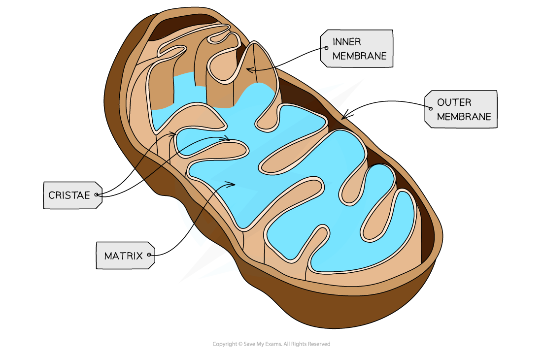
A single mitochondrion is shown – the inner membrane has protein complexes vital for the later stages of aerobic respiration embedded within it
- The site of aerobic respiration within all eukaryotic cells, mitochondria are just visible with a light microscope
- Surrounded by double-membrane with the inner membrane folded to form cristae
- The matrix formed by the cristae contains enzymes needed for aerobic respiration, producing ATP
- Small circular pieces of DNA (mitochondrial DNA) and ribosomes are also found in the matrix (needed for replication)
Golgi apparatus
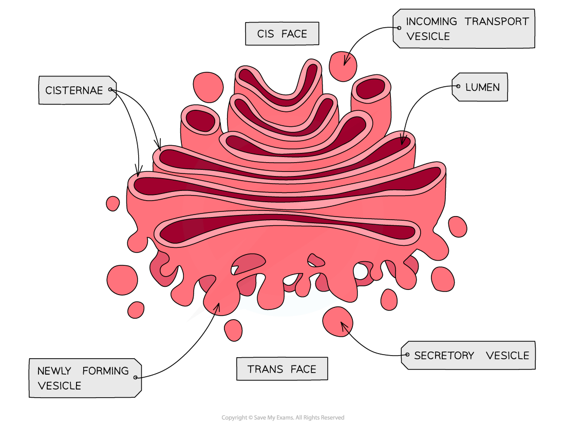
The structure of the Golgi apparatus
- Found in plant and animal cells
- Flattened sacs of membrane called cisternae (like the rough endoplasmic reticulum)
- Modifies proteins and lipids before packaging them into Golgi vesicles
- The vesicles then transport the proteins and lipids to their required destination
- Proteins that go through the Golgi apparatus are usually exported (e.g. hormones such as insulin), put into lysosomes (such as hydrolytic enzymes) or delivered to membrane-bound organelles
Vesicles
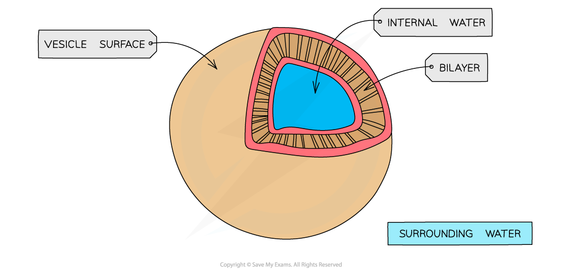
The structure of the vesicle
- Found in plant and animal cells
- A membrane-bound sac for transport and storage
Lysosome
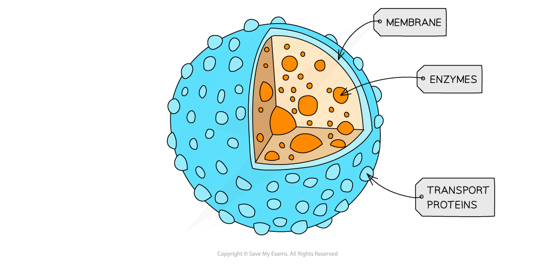
The structure of the lysosome
- Specialist forms of vesicles which contain hydrolytic enzymes (enzymes that break biological molecules down)
- Break down waste materials such as worn-out organelles
- Used extensively by cells of the immune system and in apoptosis (programmed cell death)
Chloroplasts
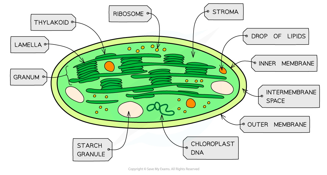
Chloroplasts are found in the green parts of a plant – the green colour a result of the photosynthetic pigment chlorophyll
- Found in plant cells
- Larger than mitochondria
- Surrounded by a double-membrane
- Membrane-bound compartments called thylakoids containing chlorophyll stack to form structures called grana
- Grana are joined together by lamellae (thin and flat thylakoid membranes)
- Chloroplasts are the site of photosynthesis:
- The light-dependent stage takes place in the thylakoids
- The light-independent stage (Calvin Cycle) takes place in the stroma
- Also contain small circular pieces of DNA and ribosomes used to synthesise proteins needed in chloroplast replication and photosynthesis
Large permanent vacuoles
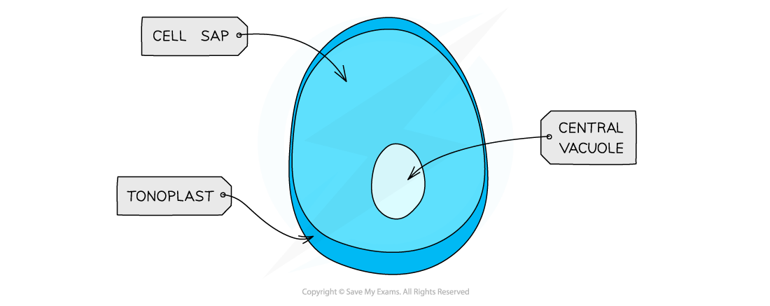
The structure of the vacuole
- A sac in plant cells surrounded by the tonoplast, selectively permeable membrane
- Vacuoles in animal cells are not permanent and small
Cell wall - an extra-cellular component (not an organelle)
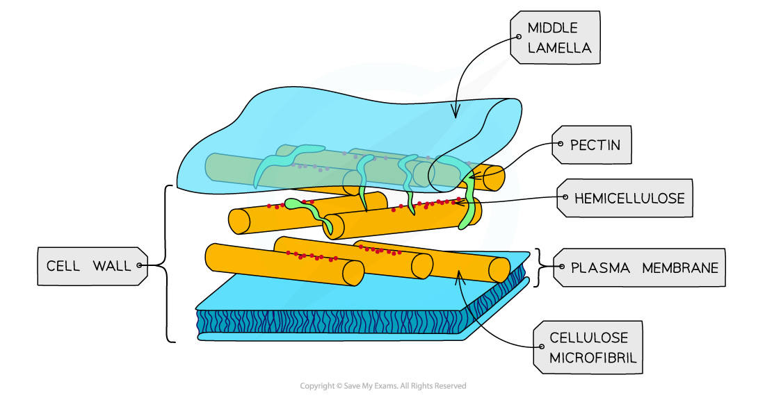
The cell wall is freely permeable to most substances (unlike the plasma membrane)
- Found in plant cells but not in animal cells
- Cell walls are formed outside of the cell membrane and offer structural support to cell
- Structural support is provided by the polysaccharide cellulose in plants, and peptidoglycan in most bacterial cells
- Narrow threads of cytoplasm (surrounded by a cell membrane) called plasmodesmata connect the cytoplasm of neighbouring plant cells
Additional organelles
- The below organelles can be found in other specialised cells in eukaryotes
Flagella

The structure of the flagella
- Found in specialised cells
- Similar in structure to cilia, made of longer microtubules
- Contract to provide cell movement for example in sperm cells
Centrioles
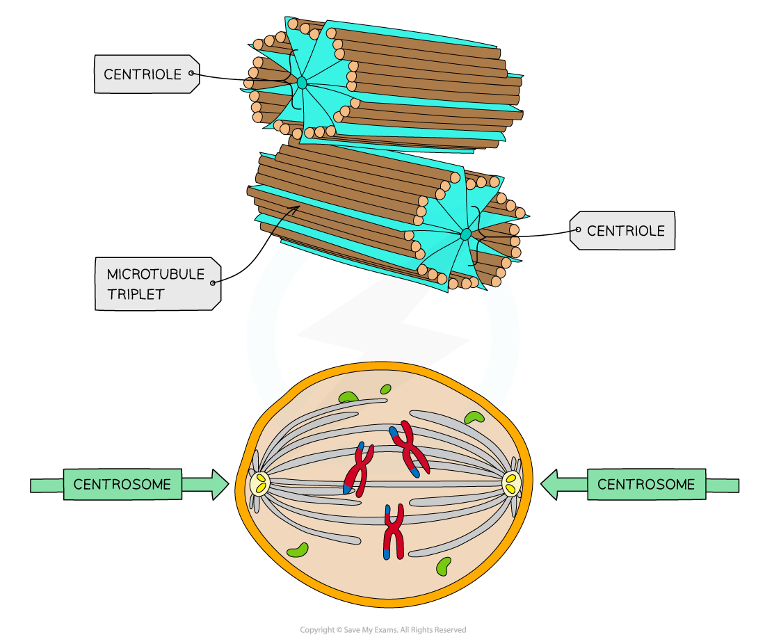
The structure of the centriole
- Hollow fibres made of microtubules
- Two centrioles at right angles to each other form a centrosome, which organises the spindle fibres during cell division
- Not found in flowering plants and fungi
Microtubules
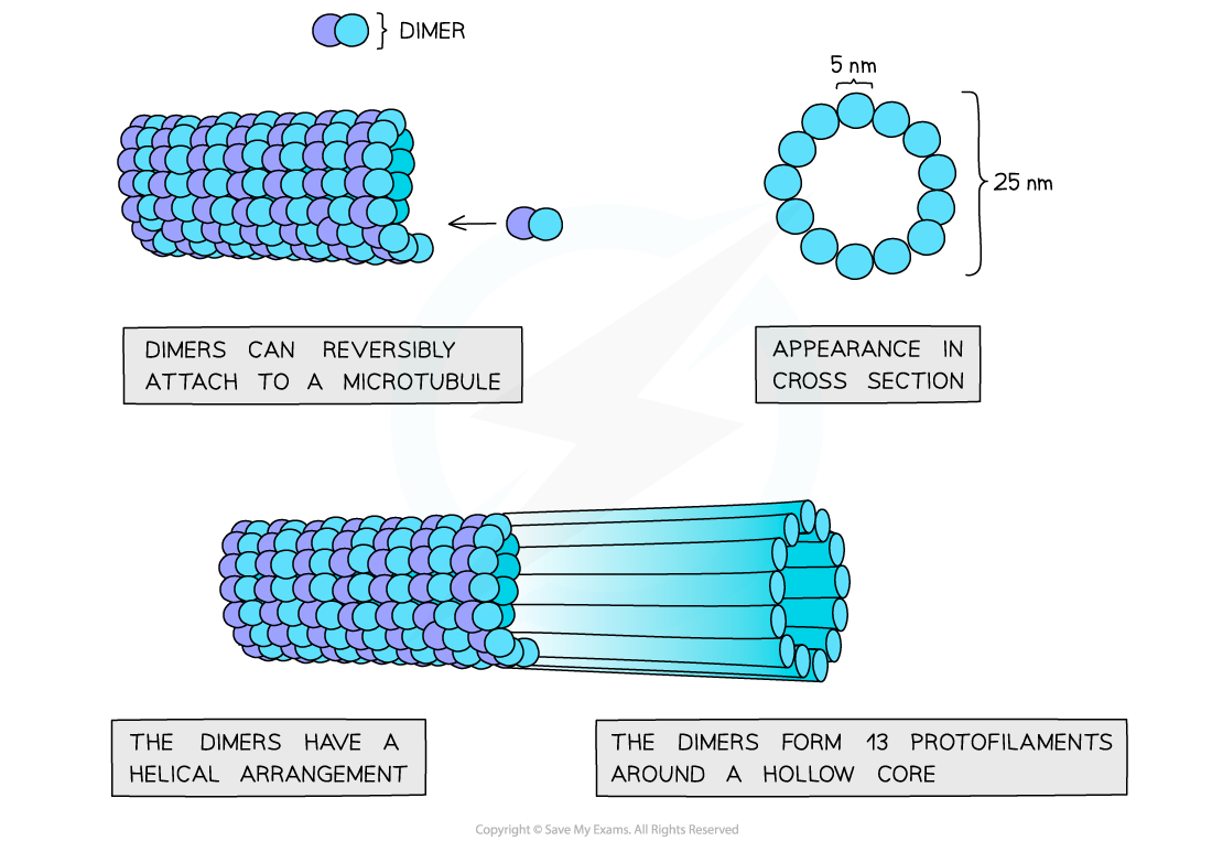
The structure of the microtubule
- Found in all eukaryotic cells
- Makes up the cytoskeleton of the cell about 25 nm in diameter
- Made of α and β tubulin combined to form dimers, the dimers are then joined into protofilaments
- Thirteen protofilaments in a cylinder make a microtubule
- The cytoskeleton is used to provide support and movement of the cell
Microvilli
The structure of the microvilli
- Found in specialised animal cells
- Cell membrane projections
- Used to increase the surface area of the cell surface membrane in order to increase the rate of exchange of substances
Cilia
The structure of the cilia
- Hair-like projections made from microtubules
- Allows the movement of substances over the cell surface
Exam Tip
In the exam, you could be required to apply your knowledge of organelles to deduce the function of a specialised cell. To answer these questions, just think about what organelles you can see in large numbers, consider the function of that organelle and then think about where this function might need to happen a lot in an organism (e.g. if the cell’s main function is to carry out photosynthesis it will need to contain many chloroplasts)!
转载自savemyexams
以上就是关于【IB DP Biology: SL复习笔记1.2.4 Eukaryotic Cell Structure】的解答,如需了解学校/赛事/课程动态,可至翰林教育官网获取更多信息。
往期文章阅读推荐:
深耕九载!30+国际竞赛/课程讲义,硕博100%团队操刀,助力爬藤冲G5!

翰林AMC8视频课重磅上线!

国际竞赛真题资源免费领取