IB DP Biology: HL复习笔记1.2.8 Skills: Drawing Cells

Drawing Cells
Drawing the ultrastructure of cells
- To record the observations seen under the microscope (or from photomicrographs taken) a labelled biological drawing is often made
- Biological drawings are line pictures that show specific features that have been observed when the specimen was viewed
- There are a number of rules/conventions that are followed when making a biological drawing
Drawing conventions
- The drawing must have a title
- The magnification under which the observations shown by the drawing are made must be recorded
- A sharp HB pencil should be used (and a good eraser!)
- Drawings should be on plain white paper
- Lines should be clear, single lines (no thick shading)
- No shading
- The drawing should take up as much of the space on the page as possible
- Well-defined structures should be drawn
- The drawing should be made with proper proportions
- Label lines should not cross or have arrowheads and should connect directly to the part of the drawing being labelled
- Label lines should be kept to one side of the drawing (in parallel to the top of the page) and drawn with a ruler
- Drawings of cells are typically made when visualizing cells at a higher magnification power, whereas plan drawings are typically made of tissues viewed under lower magnifications (individual cells are never drawn in a plan diagram)
Drawing Prokaryotic Cells
- Due to the size of prokaryotes (0.1 to 5 µm) their ultrastructure can only be seen using an electron microscope
- Therefore drawings of prokaryotes are based on electron micrographs
- When viewing an electron micrograph of a prokaryote there is no distinct dark circular area within the cell, as there is no nucleus and no organelles are visible (apart from ribosomes, but as they are 70 S in size these are difficult to distinguish)
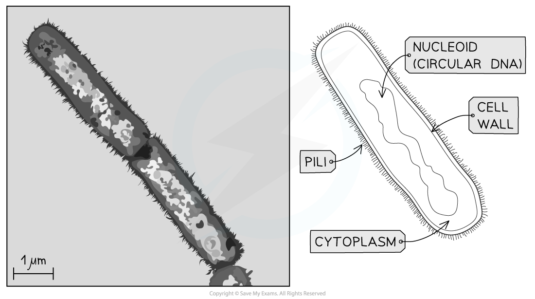
Transmission electron micrograph of a prokaryote and drawing
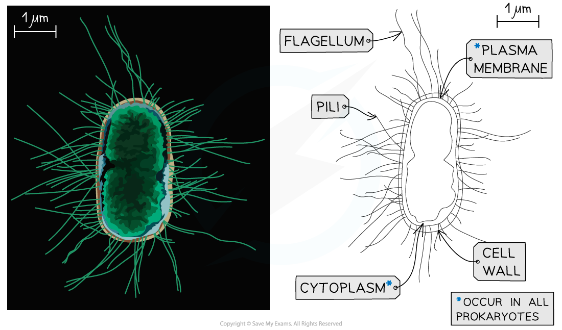
Scanning electron micrograph of an E.coli and drawing
Drawing Eukaryotic Cells
- When viewing a eukaryotic cell under a light microscope it is possible to identify the nucleus and if it is a plant cell the cell wall and vacuole
- However, under an electron microscope, more detail of the ultrastructure of the eukaryotic cell can be seen
- The following organelles should be able to be identified, although it does depend on whether it is a plant or animal cell and the specialisation of the cell:
- Rough endoplasmic reticulum
- Golgi apparatus
- Lysosomes
- Vesicles
- Ribosomes
- Vacuole (plant)
- Nucleus
- Mitochondrion
- Chloroplast
- The nucleus, mitochondrion and chloroplast all have double membranes
- The cell wall will be present in plant eukaryotic cells. This is an extra-cellular component
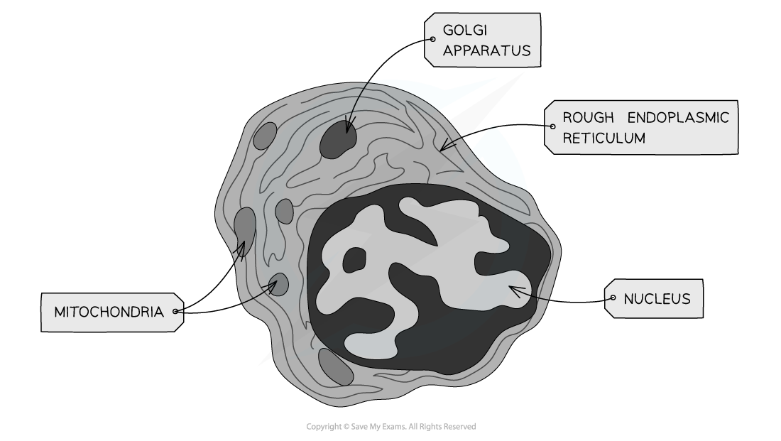
TEM electron micrograph of an animal cell showing key features
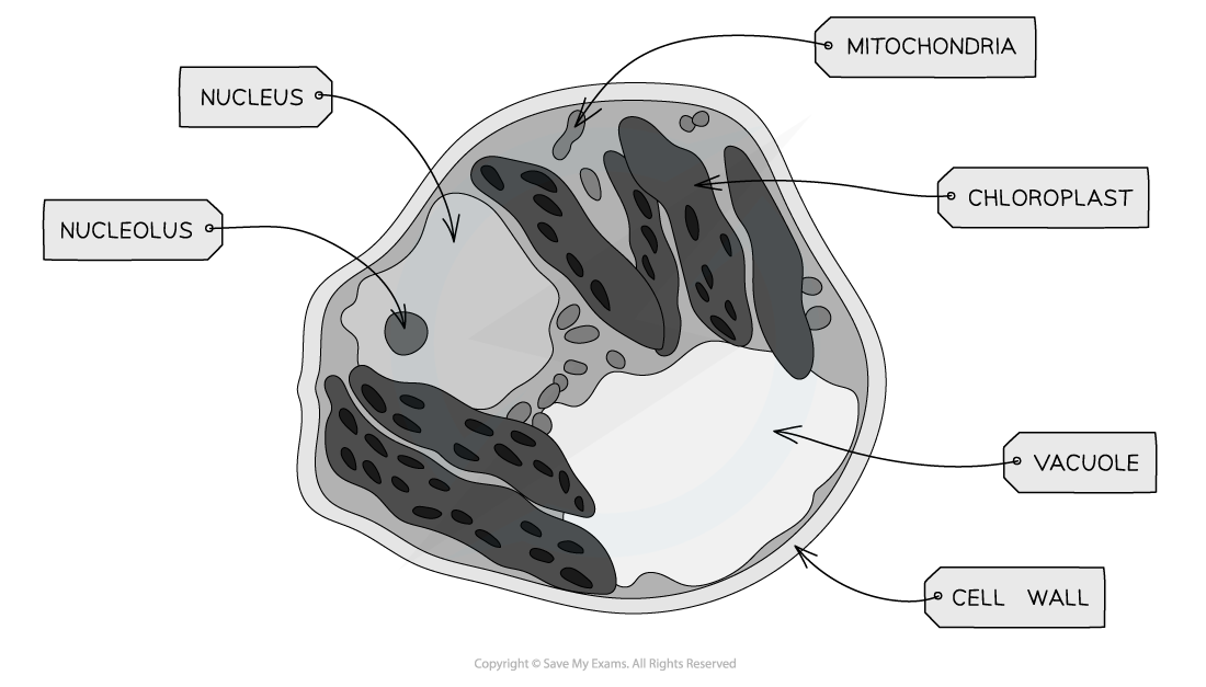
TEM electron micrograph of a plant cell showing key features
- Electron microscopes can produce highly detailed images of animal and plant cells
- The key cellular structures within animal and plant cells are visible within the electron micrographs above
- The presence of a vacuole in a micrograph is a good indicator of the cell type
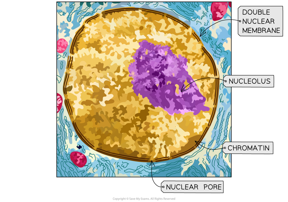
Electron micrograph of the nucleus
- The nucleus should be clearly identifiable as it is the largest structure in the eukaryotic cell
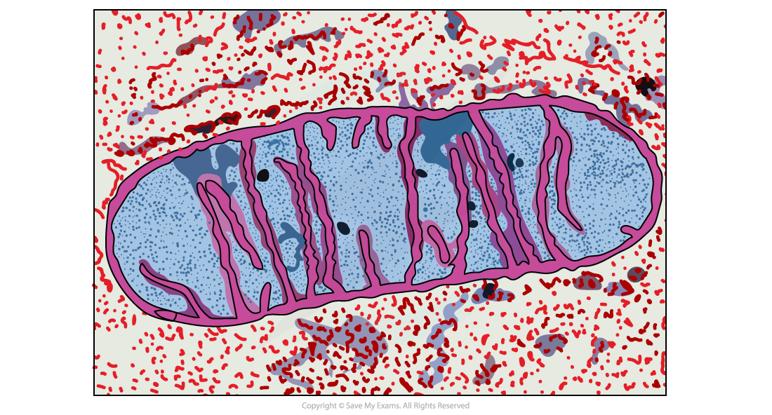
Electron micrograph of the mitochondrion
- To identify the mitochondrion look for the crista (the foldings of the inner membrane) which are often visible in electron micrographs
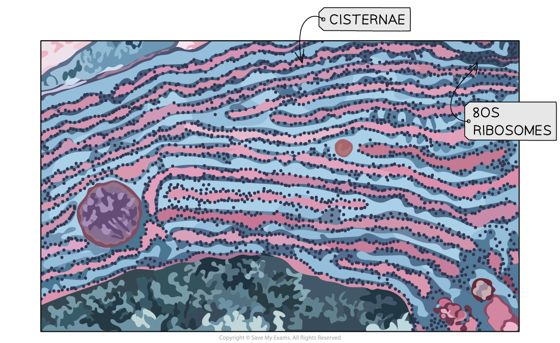
Electron micrograph of the rough endoplasmic reticulum
- The rough endoplasmic reticulum (rER) is located next to the nucleus and the attached ribosomes can be used to identify the rER as they make the membrane appear darker
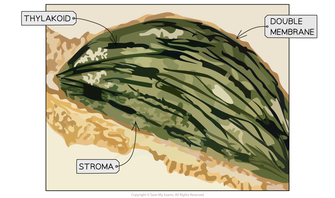
Electron micrograph of the chloroplast
- The chloroplast can be identified by the thylakoid stacks (grana), as they appear as dark lines within the organelle
- Chloroplasts are large
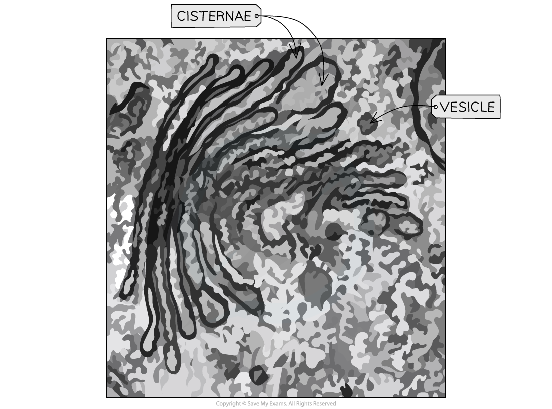
Electron micrograph of the Golgi apparatus
- Golgi apparatus will be located near the endoplasmic reticulum and it:
- Does not have long membrane sacs
- The sacs are more curved than the endoplasmic reticulum
- Does not have ribosomes attached
- Has many vesicles close by
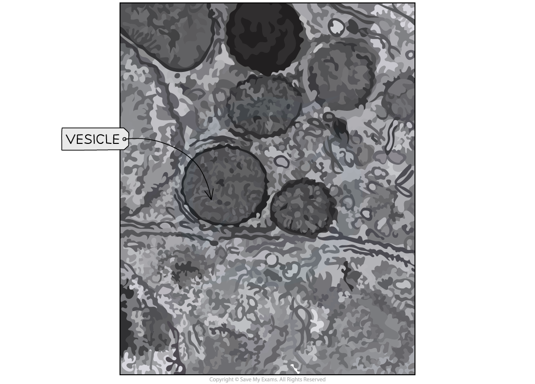
Electron micrograph of the vesicles
- Vesicles are spherical shapes
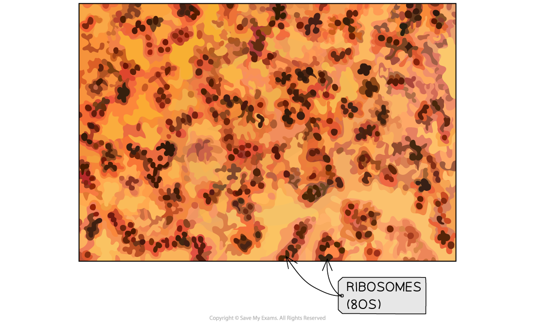
Electron micrograph of the ribosomes
- Free ribosomes appear as dark granules (tiny dark dots) in the cytoplasm
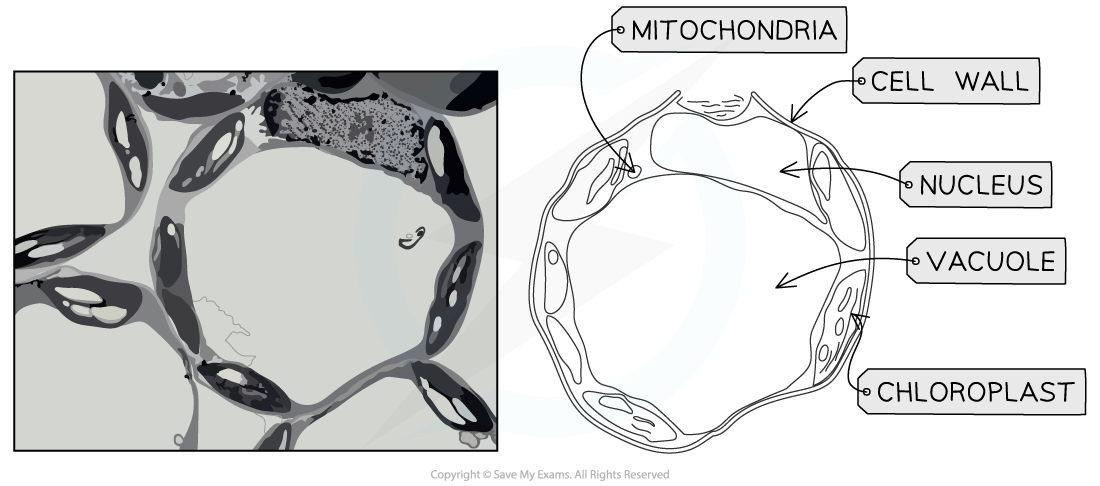
Electron micrograph and drawing of a palisade mesophyll cell
- The palisade mesophyll electron micrograph will have:
- The chloroplasts along the plasma membrane as this is where the most light can be absorbed
- A large vacuole in the centre
- A cell wall
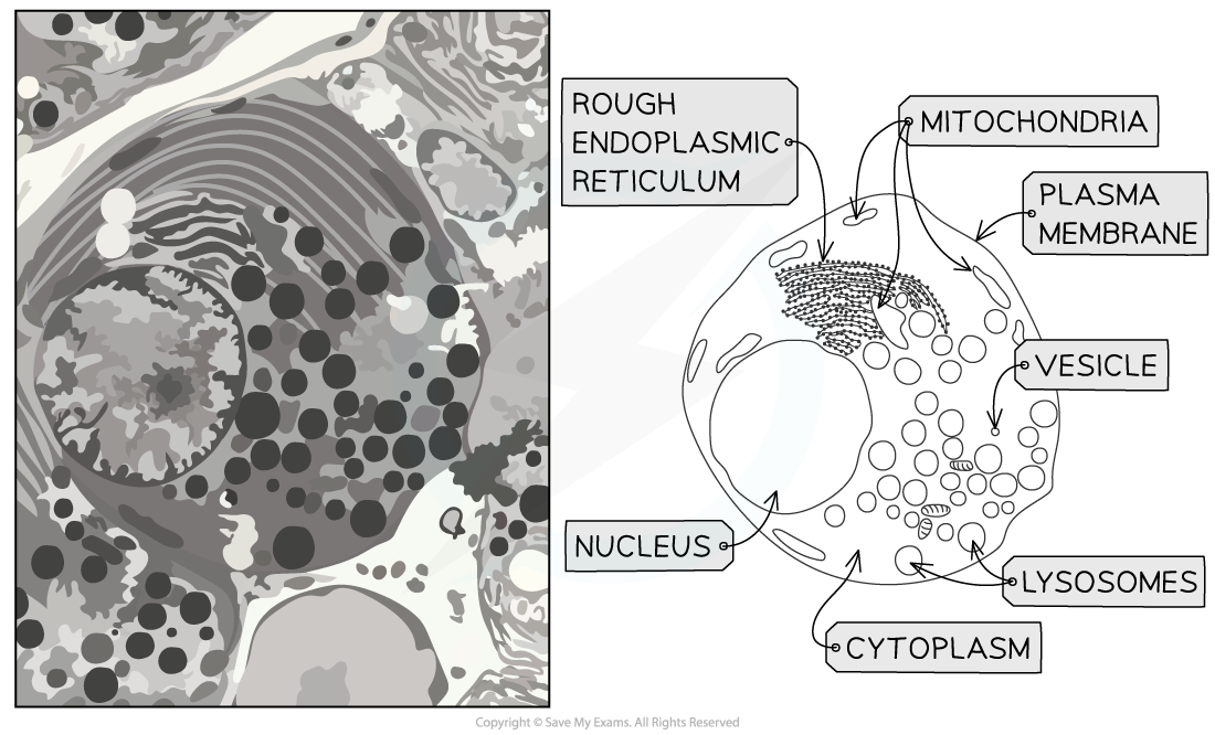
Electron micrograph and drawing of an exocrine gland cell of the pancreas
- An exocrine gland cell of the pancreas will:
- Have many large secretory vesicles (carrying the digestive enzymes)
- Have many mitochondria
- Be densely packed with rough endoplasmic reticulum
Exam Tip
When producing a biological drawing, it is vital that you only ever draw what you see and not what you think you see.When identifying the palisade mesophyll cell look for the presence of the large central vacuole, cell wall and lots of chloroplasts on the edge of the cell to maximise light absorption.When identifying the exocrine pancreatic gland cell look for the presence of secretory vesicles carrying the digestive enzymes and the large numbers of rough endoplasmic reticulum.
转载自savemyexams
以上就是关于【IB DP Biology: HL复习笔记1.2.8 Skills: Drawing Cells】的解答,如需了解学校/赛事/课程动态,可至翰林教育官网获取更多信息。
往期文章阅读推荐:
翰林独家 | 经济学竞赛核心精讲,一册打通NEC/IEO/USAEBO!
2026 AMC10/12美国数学竞赛新赛季!【翰林教育 × 清华大学出版社】独家教材全面发售!

翰林AMC8视频课重磅上线!

国际竞赛真题资源免费领取